When a pet presents with seizures, sudden paralysis, or unexplained pain, identifying the underlying cause often requires more than a physical examination and standard X-rays. Neurological conditions involve structures deep within the skull and spinal column that cannot be adequately visualised with conventional diagnostics.
Modern imaging technology has transformed the way veterinary specialists diagnose and treat these complex conditions. By providing detailed views of the brain, spinal cord, and surrounding tissues, advanced imaging allows neurologists to pinpoint problems that would otherwise remain invisible.
The Limitations of Traditional Imaging
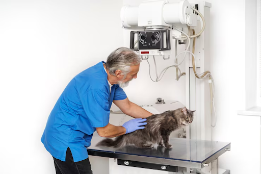
Standard radiographs are valuable for evaluating bones, joints, and organ size. However, they have significant limitations when it comes to neurological assessment. X-rays cannot visualise the spinal cord itself, detect soft tissue tumours, or reveal the subtle changes in brain structure that cause seizures or behavioural changes.
For decades, veterinarians had to rely on clinical signs and process of elimination when diagnosing neurological disease. Advanced veterinary medical imaging has changed that entirely, providing the diagnostic clarity needed to make confident treatment decisions.
MRI: The Gold Standard for Neurological Diagnosis
Magnetic resonance imaging uses powerful magnetic fields and radio waves to create detailed cross-sectional images of soft tissues. In veterinary neurology, MRI is the primary tool for evaluating the brain, spinal cord, intervertebral discs, and nerve roots.
MRI can detect brain tumours, inflammatory conditions, congenital malformations, disc herniations, and areas of spinal cord compression with a level of detail that no other imaging modality can match. The images allow the neurologist to determine the exact location, size, and nature of the problem before deciding on a treatment approach.
CT Scanning: Speed and Precision
Computed tomography provides rapid, high-resolution images of bony structures and is particularly useful for evaluating the spine, skull, and inner ear. Advanced veterinary imaging with CT is often the first choice for emergency cases where speed is critical, such as acute disc herniations or spinal fractures after trauma advanced veterinary imaging
CT scans can also be used for guided biopsies, allowing neurologists to collect tissue samples from deep brain lesions with minimal invasiveness. This capability has expanded the range of conditions that can be diagnosed without open surgery.
Electrodiagnostics: Testing Nerve and Muscle Function
Veterinary imaging services at specialised neurology centres often include electrodiagnostic testing. Electromyography measures the electrical activity in muscles, while nerve conduction studies assess how well signals travel along peripheral nerves.
These tests are essential for diagnosing conditions like peripheral neuropathies, myasthenia gravis, and nerve root disorders that may not be visible on MRI or CT. They help differentiate between nerve, muscle, and spinal cord disease when symptoms overlap.
How Imaging Guides Treatment Decisions
The value of advanced imaging extends beyond diagnosis. It directly shapes treatment planning. A surgeon reviewing an MRI can determine the exact surgical approach, estimate the extent of tissue involvement, and anticipate potential complications before entering the operating room.
For non-surgical cases, imaging helps neurologists select the most appropriate medications, monitor disease progression over time, and evaluate response to treatment through follow-up scans.
What Pet Owners Should Know
Advanced imaging in animals requires general anaesthesia to ensure the patient remains completely still during the scan. This is standard practice and is supervised by a veterinary anaesthesiologist or experienced veterinary team. The risk of anaesthesia is carefully managed and is outweighed by the diagnostic value of the imaging.
Most imaging appointments include the consultation, the scan itself, and a same-day review of results with the pet owner. In many cases, a treatment plan can be discussed within hours of the initial appointment.
Conclusion
Advanced imaging has fundamentally changed the landscape of veterinary neurology. MRI, CT, and electrodiagnostic testing give specialists the ability to see what was previously invisible, diagnose conditions accurately, and plan treatments with precision. For pets experiencing neurological symptoms, access to these technologies can make the difference between an uncertain prognosis and a clear path forward.