In Maryland, cancer is one of the leading causes of illness and death in dogs and cats, particularly as they age. The good news is that early diagnosis can significantly improve treatment outcomes and quality of life. One of the most valuable tools in identifying cancer at its earliest stages is veterinary radiology. Through advanced imaging techniques, veterinarians can detect tumors, assess disease progression, and develop effective treatment plans before symptoms become severe.
In this article, we’ll explore how veterinary radiology helps with the early detection of cancer in pets and why timely imaging can make a life-saving difference.
Understanding Veterinary Radiology
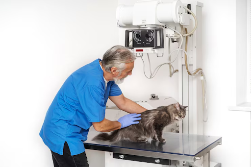
Veterinary radiology is a specialized branch of veterinary medicine that uses imaging technology to diagnose diseases and injuries in animals. It allows veterinarians to visualize internal organs, bones, and soft tissues without invasive procedures.
Common imaging techniques used in veterinary radiology include:
- X-rays (Radiographs)
- Ultrasound
- Computed Tomography (CT) scans
- Magnetic Resonance Imaging (MRI)
- Fluoroscopy
- Nuclear medicine imaging
These technologies provide detailed information that helps veterinarians identify abnormalities, including cancerous growths, at an early stage.
Why Early Cancer Detection Matters
Cancer often develops silently. By the time physical symptoms become noticeable, the disease may have already spread to other parts of the body.
Early detection offers several advantages:
- More treatment options are available.
- Tumors are generally smaller and easier to remove.
- Treatment success rates are higher.
- Pets experience a better quality of life.
- Survival times can be significantly improved.
Advanced imaging plays a crucial role in finding cancers before they become advanced or life-threatening.
Common Types of Cancer Found in Pets
Dogs and cats can develop many forms of cancer. Some of the most common include:
Lymphoma
This cancer affects the lymphatic system and can spread rapidly throughout the body.
Mast Cell Tumors
Common in dogs, these skin tumors vary in severity and often require imaging to determine whether they have spread.
Osteosarcoma
A type of bone cancer frequently seen in large-breed dogs.
Liver and Spleen Tumors
These tumors may remain hidden until they become large enough to cause internal bleeding or organ dysfunction.
Lung Cancer
Primary lung tumors are uncommon, but cancer from other areas often spreads to the lungs.
Brain Tumors
These can cause neurological symptoms such as seizures, behavioral changes, and loss of coordination.
Veterinary radiology in Maryland helps identify these cancers and determine their extent.
How X-Rays Help Detect Cancer
X-rays are often the first imaging test performed when cancer is suspected.
They are especially useful for evaluating:
- Bone tumors
- Lung masses
- Chest abnormalities
- Enlarged lymph nodes
- Abdominal masses
For example, chest radiographs are commonly used to determine whether a tumor has spread to the lungs. Early detection of metastasis helps veterinarians choose the most appropriate treatment strategy.
Although X-rays provide valuable information, some tumors are too small or located in areas that require more advanced imaging.
The Importance of Ultrasound in Cancer Diagnosis
Ultrasound uses sound waves to create real-time images of internal organs and soft tissues.
It is commonly used to examine:
- Liver
- Spleen
- Kidneys
- Bladder
- Intestinal tract
- Lymph nodes
Ultrasound can reveal abnormalities that may not appear on traditional X-rays. It also allows veterinarians to guide needle biopsies safely and accurately.
For pets with unexplained weight loss, vomiting, or abdominal swelling, ultrasound often provides crucial clues that lead to an early cancer diagnosis.
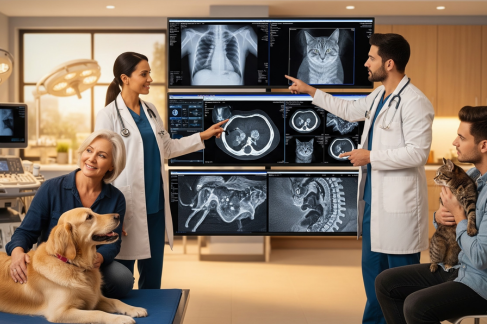
CT Scans Provide Detailed Cross-Sectional Images
Computed Tomography (CT) scans combine multiple X-ray images to create highly detailed cross-sectional views of the body.
Veterinary CT scans in Maryland are particularly valuable for:
- Detecting nasal tumors
- Evaluating lung cancer
- Identifying bone tumors
- Assessing tumors in the chest and abdomen
- Planning surgical procedures
Because CT imaging produces three-dimensional views, veterinarians can determine the size and location of tumors with remarkable accuracy.
This information is essential when deciding whether surgery, radiation therapy, or other treatments are appropriate.
MRI Helps Diagnose Brain and Spinal Tumors
Magnetic Resonance Imaging (MRI) provides exceptional detail of soft tissues and nervous system structures.
Veterinary MRI is considered the gold standard for diagnosing:
- Brain tumors
- Spinal cord tumors
- Nerve sheath tumors
- Soft tissue cancers
Pets experiencing seizures, head tilt, weakness, or unexplained neurological symptoms often benefit from MRI examinations.
Early identification of tumors affecting the brain or spinal cord can dramatically improve treatment outcomes and help preserve neurological function.
Staging Cancer Through Advanced Imaging
Finding a tumor is only the first step. Veterinary radiology is also essential for cancer staging, which determines how far the disease has progressed.
Imaging helps veterinarians assess:
- Tumor size
- Involvement of nearby tissues
- Lymph node enlargement
- Spread to distant organs
- Surgical feasibility
Accurate staging allows veterinary oncologists to design personalized treatment plans and provide realistic prognoses.
Monitoring Treatment Progress
Veterinary radiology remains important even after a cancer diagnosis.
Imaging is frequently used to:
- Measure tumor response to chemotherapy
- Monitor radiation therapy effectiveness
- Detect recurrence
- Identify complications
- Evaluate post-surgical healing
Regular imaging enables veterinarians to adjust treatment strategies when necessary and ensure pets receive the best possible care.
Which Pets May Need Cancer Screening?
Certain pets are at higher risk for cancer and may benefit from routine imaging evaluations.
Risk factors include:
- Senior age
- Large-breed dogs
- Previous cancer diagnosis
- Family history of cancer
- Persistent unexplained symptoms
- Abnormal blood test results
Breeds commonly predisposed to cancer include:
- Golden Retrievers
- Boxers
- Bernese Mountain Dogs
- German Shepherds
- Labrador Retrievers
For these pets, early screening through veterinary radiology can detect problems before symptoms become severe.
Benefits of Veterinary Radiology in Cancer Care
Modern imaging technology offers several advantages:
- Non-invasive diagnosis
- Earlier detection of tumors
- Improved treatment planning
- Better surgical outcomes
- More accurate cancer staging
- Enhanced monitoring during treatment
- Increased quality of life for pets
Advances in veterinary radiology continue to transform cancer care, giving pets access to more precise and effective treatment options than ever before.
Final Thoughts
Cancer is a serious diagnosis, but early detection can make a tremendous difference in a pet’s prognosis. Veterinary radiology plays a vital role in identifying tumors, determining disease extent, and guiding treatment decisions. From X-rays and ultrasound to CT scans and MRI, advanced imaging technologies help veterinarians detect cancer sooner and provide pets with the best chance for successful treatment.
If your dog or cat is experiencing unusual symptoms or belongs to a breed with an increased risk of cancer, discussing diagnostic imaging with your veterinarian may be one of the most important steps you can take to protect their health and well-being.